1- Disruption of thyroid follicles , with extravasation of colloid leading to a PMN infiltrate.
2- subacute granulomatous thyroiditis
3- viral infection
2- subacute granulomatous thyroiditis
3- viral infection
HODGKIN LYMPHOMA
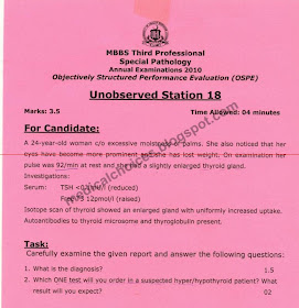
graves disease
TSH
TSH
1. bronchioalveolar carcinoma 2. lepidic growth which grows along pre existing structures without distorting the alveoli 3. muscle weakness due to antibodies against neuronal calcium channels
obstructive jaundice,,alk po4ase raised,ast alt,albumin
Fibroadenoma
types are giant , intracanlicular n pericanalicular
formalin
MPGN
Immunofluorescence staining
three types : Minimal change, rapid progressive n membranous
3 types are tubular , villous n tubulovillous
 |
| 1.Protein loss greater than 3.5 g in nephrotic |
1 ) -single layer of Tall columnar epithelium , they are ciliated n some part are dome shaped secretary cells .
- Psommoma bodies
2) Serous type
3) Mucinous n endometroid
| Infarct 4 factors: occlusion of an artery (atherosclerosis) , thrombosis, embolus, smoking, age, sex, etc 3. 5hrs-early coagulative necrosis, edema, hemorrhage |
1. lesser curvature, 2. gastric ca confined to mucosa and submucosa regardless of the mets, 3. gastric ca met subcutaneous nodule in the periumbilical region
 |
| 1.Diabetic ketoacidosis2. metabloic acidosis |
 |
| bronchopneumonia |
 |
| leiomyoma |
1- seminoma cells : large , round to polyhydral cells wth distinct cell membranes , clear cytoplasm n central round nuclei with infrequent mitosis
2- Seminoma
3- Non differentiated totipotential germ cell tumor
Polcythemia vera
bone marrow biopsy n level will b low
1- Gaint cell tumor of bone
2- Malignant
3- GCTs are large, purely lytic n eccentric, the overall cortex is destroyed producing a bulging soft tissue mass.
4-Reactive macrophages n mononuclear celss
 |
| CML |
1- malignant cells disrupting the norml epidermal barrier
2- paget disease of nipple
3- DCIS
peptic ulcer diagnoses, syndrome zollinger ellison syndrome complication: bleeding, perforation, obstruction
 |
| GIANT CELL ARTEITIS |
restrictive lung diseases"honeycomb"
1- cut surface shows fibrosis ( firm, rubbery white areas)
2- Idiopathic pulmonary fibrosis n pneumoconiosis
3-Interstitium in the alveolar wall
Ulcerative colitis : The pale irregular regions comprise ulcerations, n there is also pseudopolyp formations.
UC involve rectum , sigmoid n may involve entire colon
post-infectious glomerulonephritis
1. proliferation of mesingium, epithelial cells and infiltration with neutrophils...
2. Streptococcus with M protein
3. INCREASED ASO TITER, rbc cast in urine
2. Streptococcus with M protein
3. INCREASED ASO TITER, rbc cast in urine
| Iron deficency anemia causes : low dietry intake , malabsorption n increase demand or chronic blood loss |
 |
| meningioma |
 |
| papillary carcinoma |
 |
| pernicious anemia |
 |
| "clear cell carcinomas" chromophobe has bst prognosis . |


No comments:
Post a Comment